德晋贵宾厅

B-Tg(Luc-EGFP) PANC-1

NA • 322436

B-Tg(Luc-EGFP) PANC-1

Product nameB-Tg(Luc-EGFP) PANC-1
Catalog number322436
Strain nameNA

在此页面上

  • Description
  • Efficacy

海报

查看全部

    发表文章

      Description
      • Origin: PANC-1 is an epithelial morphology cell line, established from a pancreatic duct specimen obtained from a 56-year-old, White, male patient diagnosed with epithelioid carcinoma. This cell line can be used in cancer research.
      • Background Information: Luciferase-expressing cell lines are genetically engineered to produce luciferase enzymes, typically from fireflies (Photinus pyralis). These bioluminescent reporter systems enable real-time monitoring of cellular processes through light emission when exposed to substrate luciferin. Widely used in molecular biology, they facilitate gene expression studies, drug screening, and in vivo imaging applications. EGFP provides a stable fluorescent marker for reliable visualization and tracking of tumor cells, enabling convenient monitoring of cell growth and tumor progression.
      • Gene targeting strategy: The exogenous promoter and luciferase-P2A-EGFP coding sequence were randomly inserted into the PANC-1 cells.
      • Tumorigenicity: Confirmed in B-NDG mice.
      • Application: The B-Tg(Luc-EGFP) PANC-1 tumor model is suitable for preclinical evaluation of antitumor efficacy and therapeutic responses using bioluminescence imaging.
      In Vitro Luciferase Activity and EGFP Expression Analysis

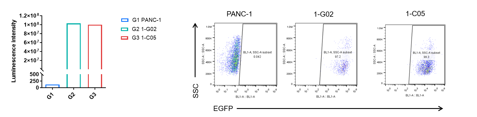
      Luminescence signal intensity of B-Tg(Luc-EGFP) PANC-1 cells and EGFP expression analysis in B-Tg(Luc-EGFP) PANC-1 cells. Single cell suspensions from wild-type PANC-1 and B-Tg(Luc-EGFP) PANC-1 #1-G02, #1-C05 cultures were measured using the Bright-GloTM luciferase Assay (Promega, Catalog No. E4030). B-Tg(Luc-EGFP) PANC-1 cells have a strong luminescence signal that is not present in wild-type PANC-1 cells. EGFP was detected on the surface of B-Tg(Luc-EGFP) PANC-1 cells but not wild-type PANC-1 cells.

      Bioluminescence Imaging

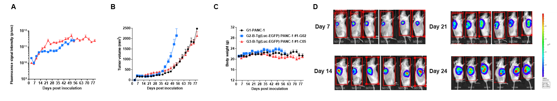
      Growth kinetics of B-Tg(Luc-EGFP) PANC-1 tumors determined by bioluminescence imaging (BLI). B-Tg(Luc-EGFP) PANC-1 cells (5×10⁶) and PANC-1 cells (5×10⁶) were subcutaneously implanted into B-NDG mice (female, n=6). Signal intensity, tumor volume and body weight were measured twice a week. (A) Signal intensity. (B) Tumor volume. (C) Body weight. (D) Raw bioluminescence images. These results indicate that B-Tg(Luc-EGFP) PANC-1 cells can be used for in vivo efficacy evaluation. Values are expressed as mean ± SEM.