C57BL/6JNifdc-Kltm1Bcgen/Bcgen • 113377
| Product name | B-Klotho KO mice |
|---|---|
| Catalog number | 113377 |
| Strain name | C57BL/6JNifdc-Kltm1Bcgen/Bcgen |
| Strain background | C57BL/6JNifdc |
| NCBI gene ID | 16591 (Mouse) |
| Aliases | alpha-kl |
Gene targeting strategy for B-Klotho KO mice.The exons 2~5 of mouse Klotho gene were knocked out in B-Klotho KO mice.
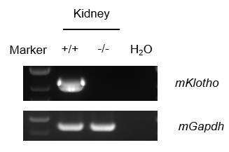
Strain specific analysis of Klotho mRNA expression in wild-type C57BL/6JNifdc mice and B-Klotho KO mice by RT-PCR. Kidney RNA were isolated from wild-type C57BL/6JNifdc mice (+/+) and homozygous B-Klotho KO mice (-/-), then cDNA libraries were synthesized by reverse transcription, followed by PCR with mouse Klotho primers. Mouse Klotho mRNA was only detectable in wild-type but not in homozygous B-Klotho KO mice.
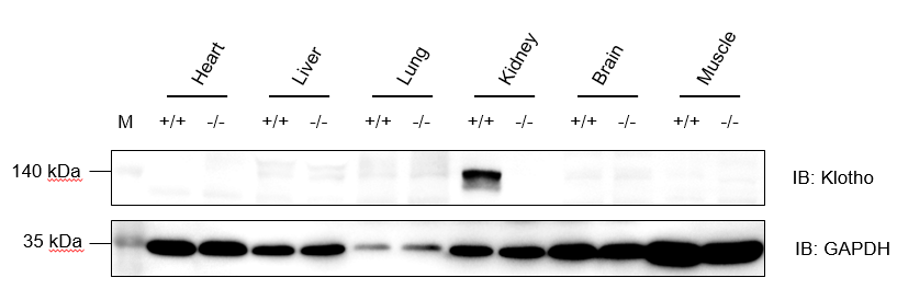
Western blot analysis of Klotho protein expression in wild-type C57BL/6JNidc mice and homozygous B-Klotho KO mice by WB. Various tissues were collected from wild-type C57BL/6JNifdc mice (+/+) and homozygous B-Klotho KO mice (-/-), and then analyzed by western blot with anti-Klotho antibody (abcam, ab181373). 50 μg total proteins were loaded for western blotting analysis. GAPDH were detected as internal control. Klotho was detectable in kidney from C57BL/6JNifdc but not in homozygous B-Klotho KO mice.
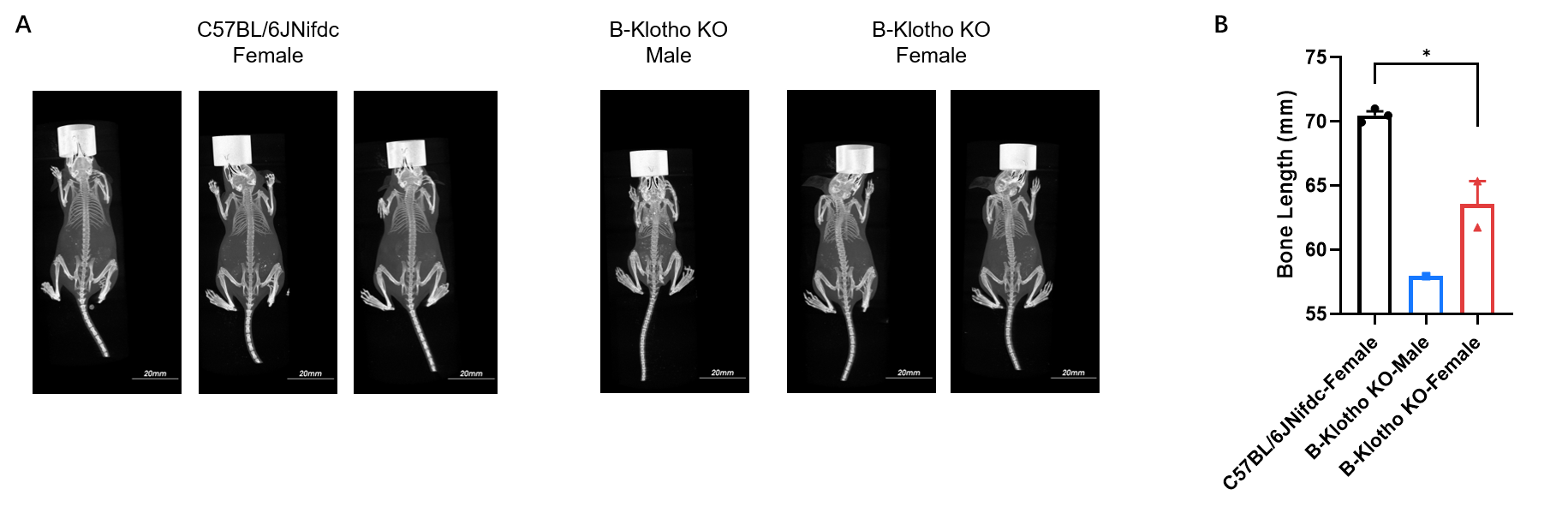
Whole-body micro-CT imaging of homozygous B-Klotho KO mice. (A) Representative whole-body micro-CT images of mice wild-type C57BL/6JNifdc mice (n=3, female, 5-week-old) and homozygous B-Klotho KO mice (n=1, male, 5-week-old; n=2, female, 5-week-old). (B) Quantitative comparison of body length (from oral-nasal region to tail tip). Body size of homozygous B-Klotho KO mice was smaller than wild-type C57BL/6JNifdc mice. Data are presented as mean ± SEM. *P < 0.05, **P < 0.01, ***P < 0.001. Scale bar = 20 mm.
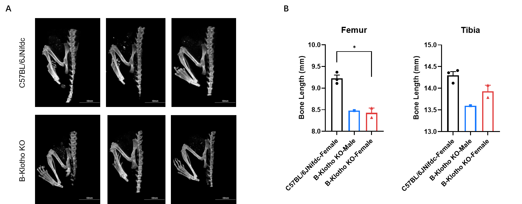
Micro-CT analysis of femur and tibia length in homozygous B-Klotho KO mice. (A) Representative micro-CT images of femur and tibia from wild-type C57BL/6JNifdc mice (n=3, female, 5-week-old) and homozygous B-Klotho KO mice (n=1, male, 5-week-old, left; n=2, female, 5-week-old, right). (B) Quantitative comparison of femur and tibia length. In the homozygous B-Klotho KO mice, the lengths of the femur and tibia were shortened. Data are presented as mean ± SEM. *P < 0.05, **P < 0.01, ***P < 0.001. Scale bar = 10 mm.
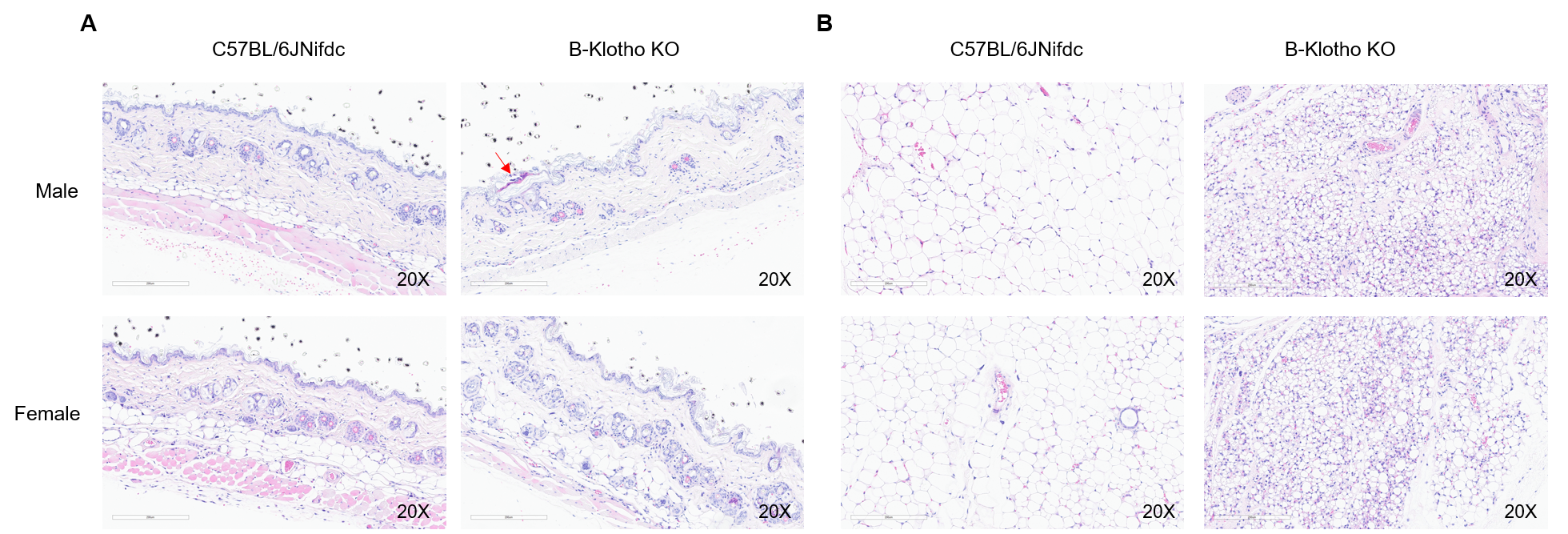
Histopathological analysis of dorsal skin and subcutaneous adipose tissue in homozygous B-Klotho KO mice. (A) H&E-stained sections showed skin pathological changes with punctate crusts, hyperkeratosis and subcutaneous fat layer was not visible in homozygous B-Klotho KO mice (-/-, 7-week-old, 1 male and 1 female) compared with wild-type C57BL/6JNifdc mice (+/+, 7-week-old, 1 male and 1 female). (B) H&E-stained subcutaneous adipose tissue showed adipocyte atrophy in homozygous B-Klotho KO mice compared with wild-type C57BL/6JNifdc. Scale bars: 200 µm.
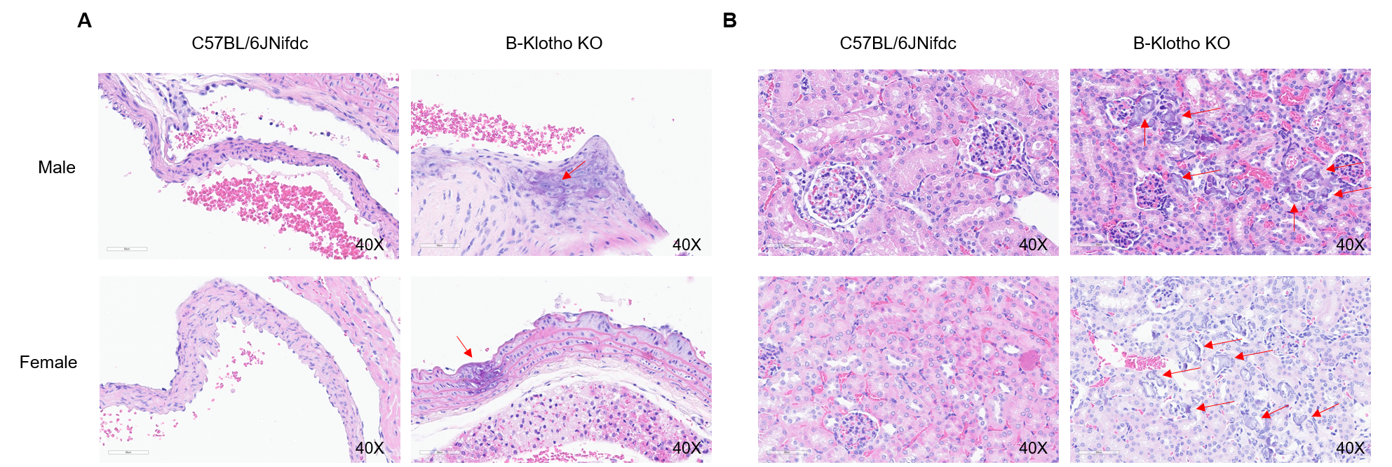
Histopathological analysis of aortas and kidney tissue in homozygous B-Klotho KO mice. H&E-stained sections showed aortas (A) and kidney (B) pathological changes with calcification of the aorta and renal vasculature in homozygous B-Klotho KO mice (-/-, 7-week-old, 1 male and 1 female) compared with wild-type C57BL/6JNifdc mice (+/+, 7-week-old, 1 male and 1 female). Scale bars: 60 µm.