
C57BL/6JNifdc-Tg(Tol2-hRHO-VEGFA)Bcgen/Bcgen • 113126
| Product name | B-Tg(hVEGFA) mice |
|---|---|
| Catalog number | 113126 |
| Strain name | C57BL/6JNifdc-Tg(Tol2-hRHO-VEGFA)Bcgen/Bcgen |
| Strain background | C57BL/6JNifdc |
| NCBI gene ID | 7422 (Human) |
| Aliases | VPF; VEGF; MVCD1; L-VEGF |
Gene targeting strategy for B-Tg(hVEGFA) mice. The exogenous human rhodopsin promoter and human VEGFA coding sequence CDS and 3’UTR were inserted into the mouse genome randomly.
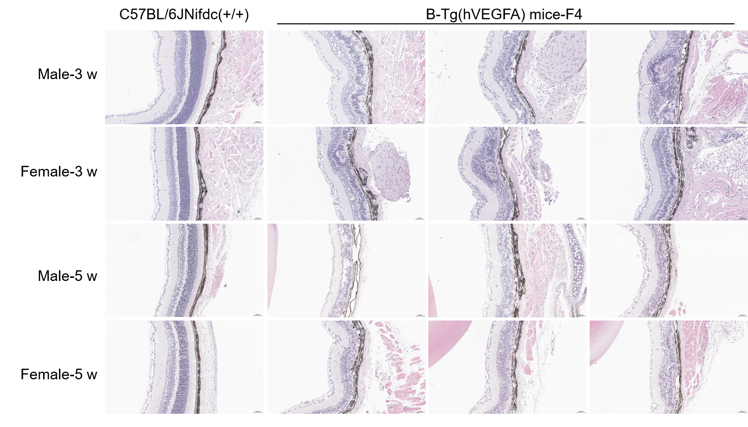
Representative images of HE staining of wild-type C57BL/6JNifdc mice and B-Tg(hVEGFA) mice. Retina tissues of wild-type C57BL/6JNifdc mice (+/+) and B-Tg(hVEGFA) mice (3 weeks old and 5 weeks old, n=3) were collected and analyzed with H&E staining. Compared with wild-type mice, B-Tg(hVEGFA) mice at 3 weeks of age displayed disorganized retinal layers (ganglion cell, inner nuclear, outer plexiform, outer nuclear) and an indistinct photoreceptor layer. By 5 weeks, the pathology worsened, with the outer nuclear layer becoming indistinct. Scale bar, 50 μm.
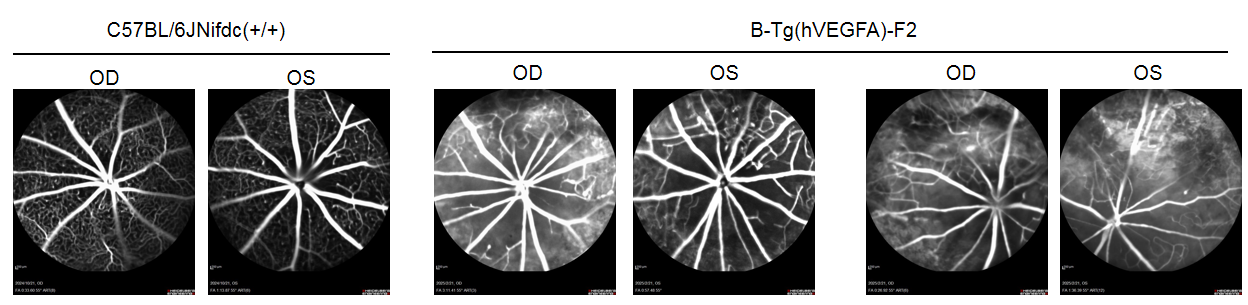
Fundus Fluorescein Angiography (FFA) of wild-type C57BL/6JNifdc mice and B-Tg(hVEGFA) mice. The FFA images of wild-type C57BL/6JNifdc mice (+/+) and B-Tg(hVEGFA) mice (12 weeks old, male). The results showed that there were uneven vascular fluorescence with patchy areas of intense fluorescence suggests vascular permeability leakage or localized pathology in B-Tg(hVEGFA) mice compared to the C57BL/6JNifdc mice.
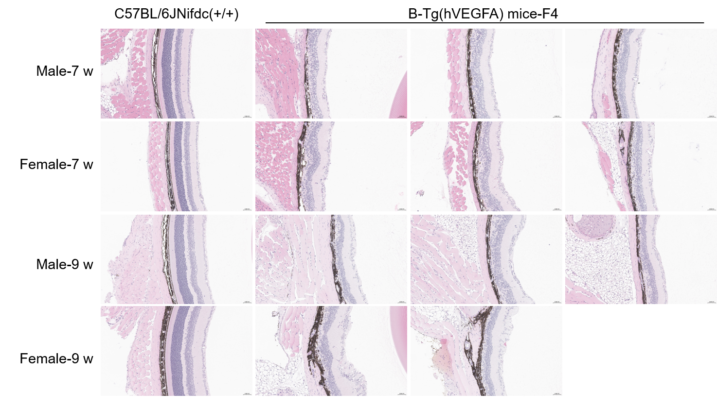
Representative images of HE staining of wild-type C57BL/6JNifdc mice and B-Tg(hVEGFA) mice. Retina tissues of wild-type C57BL/6JNifdc mice (+/+) and B-Tg(hVEGFA) mice (7 weeks old, n=3; 9 weeks old, male n=3 and female n=2) were collected and analyzed with H&E staining. The results showed that the ganglion cell layer, inner nuclear layer, outer plexiform layer, outer nuclear layer were disorganized, with the photoreceptor and outer nuclear layers being indistinct in B-Tg(hVEGFA) mice compared to the C57BL/6JNifdc mice. Scale bar, 50 μm.